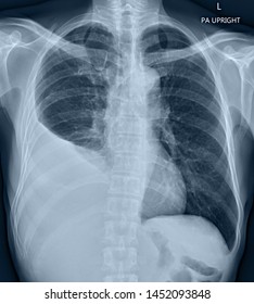
Loculated Pleural Effusion / Rul Atelectasis Iii : Malignant pleural effusion is a frequent complication of some common cancers.. Treatment may fail if the catheter is not placed optimally within the loculation or if the fluid is hemorrhagic or fibrinous. Pleural effusion is the accumulation of excess fluid in the lung space, the space between the membrane lining the lungs and the membrane lining the chest wall. Pleural effusion that is confined to one or more fixed pockets in the pleural space. A pleural effusion is due to the manifestations of another illness. Surgical thoracostomy tube placement and radiologically guided catheter drainage are standard therapy for loculated pleural fluid collections.
Of the 22 transudates, eight showed a loculated pleural effusion (36%) compared with 45 of 78 exudates (58%). An ultrasound, chest computed tomograp. The pleural fluid is an exudate that usually has predominantly lymphocytes. Empyema is defined by purulent fluid collection in the pleural space, which is most commonly caused by pneumonia. We present a unique case in which a patient presented to the ed in respiratory distress.
A Loculated Pleural Effusion A Complex Pleural Effusion Is Shown With Download Scientific Diagram from www.researchgate.net If the fluid cannot be drained, the lungs aren't able to expand and oxygenate the blood sufficiently. The pleural fluid is an exudate that usually has predominantly lymphocytes. Pleural effusion is an accumulation of fluid in the pleural space that is classified as transudate or exudate according to its composition and underlying pathophysiology. In vitro efficacy of varidase versus streptokinase or urokinase for liquefying thick purulent exudative material from loculated empyema. Tuberculous pleural effusion (tpe) is one of the most common forms of extrapulmonary tuberculosis. Loculation most commonly occurs with exudative fluid, blood and pus. Streptokinase appears to improve the resolution of loculated pleural effusions when chest tube drainage fails to achieve symptomatic relief. We report a case in which loculated recurrent pleural effusion was treated by insertion of an indwelling tenckhoff catheter.
Left pleural effusion with high density material at the posterior costophrenic angle.
Complex septated, complex nonseptated, or homogeneously echogenic effusions are always exudates (fig. This often causes shortness of breath as the lung gets compressed from the fluid. Pleural effusion that is confined to one or more fixed pockets in the pleural space. Encysted pleural fluid is visualized between the right upper and middle lobe(s). Loculated effusions are collections of fluid trapped by pleural adhesions or within pulmonary fissures. Normally, a small amount of fluid is present in the pleura. An interference in the function of fluid production or reabsorption will lead to fluid. The reasons for effusion are many, and the specific diagnosis is often based upon tap or drainage of the fluid. Pleural effusion predominantly presents with breathlessness, but cough and pleuritic chest pain can be a feature. An ultrasound, chest computed tomograp. Left pleural effusion with high density material at the posterior costophrenic angle. Of the 22 transudates, eight showed a loculated pleural effusion (36%) compared with 45 of 78 exudates (58%). The purpose of this study was to assess the value of intrapleural urokinase (uk) instillations in enhanc ing tube drainage of loculated, complex pleural effusions.
Streptokinase appears to improve the resolution of loculated pleural effusions when chest tube drainage fails to achieve symptomatic relief. Surgical thoracostomy tube placement and radiologically guided catheter drainage are standard therapy for loculated pleural fluid collections. Lung scarring and a permanent decrease in lung function are associated with chronic pleural effusion. We present a unique case in which a patient presented to the ed in respiratory distress. A pleural effusion is due to the manifestations of another illness.
Loculated Pleural Effusion Images Stock Photos Vectors Shutterstock from image.shutterstock.com The etiology of the pleural effusion determines other signs and symptoms. Of loculated pleural effusions* jeffreys. Initial testing … lupus pleuritis and other causes of pleural effusions in lupus patients. Pleural effusion is an accumulation of fluid in the pleural space that is classified as transudate or exudate according to its composition and underlying pathophysiology. Icu patients cannot sit up and the effusion layers posteriorly. Obliteration of left costophrenic angle with a wide pleural based dome shaped opacity projecting into the lung noted tracking along the cp angle and lateral chest wall suggestive of loculated pleural effusion, however the possibility of empyema can not be ruled out completely. In vitro efficacy of varidase versus streptokinase or urokinase for liquefying thick purulent exudative material from loculated empyema. Tuberculous pleural effusion (tpe) is one of the most common forms of extrapulmonary tuberculosis.
The reasons for effusion are many, and the specific diagnosis is often based upon tap or drainage of the fluid.
An interference in the function of fluid production or reabsorption will lead to fluid. Surgical treatment of pleural effusion may include chest. Tpe usually presents as an acute illness with fever, cough and pleuritic chest pain. A pleural effusion is due to the manifestations of another illness. Pleural effusion is extra fluid around the lung. The largest pocket of fluid is present posteriorly at the right lung base, with associated atelectasis and minor consolidation. Pleural effusion predominantly presents with breathlessness, but cough and pleuritic chest pain can be a feature. Empyema is defined by purulent fluid collection in the pleural space, which is most commonly caused by pneumonia. Pleural effusions describe fluid between the two layer of tissue (pleura) that cover the lung and the lining of the chest wall. Nine of the 19 malignant effusions showed loculation (47%). Obliteration of left costophrenic angle with a wide pleural based dome shaped opacity projecting into the lung noted tracking along the cp angle and lateral chest wall suggestive of loculated pleural effusion, however the possibility of empyema can not be ruled out completely. The pleural fluid is an exudate that usually has predominantly lymphocytes. The purpose of this study was to assess the value of intrapleural urokinase (uk) instillations in enhanc ing tube drainage of loculated, complex pleural effusions.
Pleural effusion is the accumulation of excess fluid in the lung space, the space between the membrane lining the lungs and the membrane lining the chest wall. A pleural effusion is due to the manifestations of another illness. Left pleural effusion with high density material at the posterior costophrenic angle. Fibrotic scar tissue may form in the pleural cavity (called loculation), preventing effective drainage of the fluid. Diffuse nodules and opacification in right lung with compressive atelectasis.
Lti 01 Lung Therapeutics from d1io3yog0oux5.cloudfront.net Pleural effusion is an accumulation of fluid in the pleural space that is classified as transudate or exudate according to its composition and underlying pathophysiology. Twenty of the 21 complicated parapneumonic effusions (including empyemas) showed loculation (95%). We report a case in which loculated recurrent pleural effusion was treated by insertion of an indwelling tenckhoff catheter. Causes of an exudative effusion are malignancy, infection, or inflammatory disorders such as rheumatoid arthritis. Obliteration of left costophrenic angle with a wide pleural based dome shaped opacity projecting into the lung noted tracking along the cp angle and lateral chest wall suggestive of loculated pleural effusion, however the possibility of empyema can not be ruled out completely. Tube thoracostomy has variable success in the treatment of complex pleural effusions, with Loculation most commonly occurs with exudative fluid, blood and pus. Pocus demonstrated a large right sided loculated pleural effusion with associated septations and surrounding consolidation suggestive of a parapneumonic effusion.
Pocus demonstrated a large right sided loculated pleural effusion with associated septations and surrounding consolidation suggestive of a parapneumonic effusion.
Pleural effusion is the accumulation of excess fluid in the lung space, the space between the membrane lining the lungs and the membrane lining the chest wall. Of loculated pleural effusions* jeffreys. 681 views reviewed >2 years ago Loculated pleural effusion the pleura is a thin membrane between the lungs and chest wall that lubricates these surfaces and allows movement of the lungs while breathing. Pleural effusion predominantly presents with breathlessness, but cough and pleuritic chest pain can be a feature. Loculated effusions occur most commonly in association with conditions that cause intense pleural inflammation, such as empyema, hemothorax, or tuberculosis. Lung scarring and a permanent decrease in lung function are associated with chronic pleural effusion. Tpe usually presents as an acute illness with fever, cough and pleuritic chest pain. Nine of the 19 malignant effusions showed loculation (47%). This often causes shortness of breath as the lung gets compressed from the fluid. If the fluid cannot be drained, the lungs aren't able to expand and oxygenate the blood sufficiently. 1 article features images from this case 20 public playlist include this case The largest pocket of fluid is present posteriorly at the right lung base, with associated atelectasis and minor consolidation.


0 Komentar